Category Archives: Microscopes and Magnification
Fruit Fly Development: Cell by Cell [Nature Video]

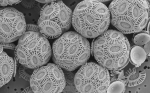
Drosophila melanogaster
Wow. Two papers published in Nature Methods have outlined a new technique which allows researchers to track development of embryos (in this case Drosophila melanogaster), in real time. By taking simulataneous multi-view microscopic images of the developing embryo, individual cells can be tracked in real time. The methods are described in more detail at Nature News here.
Have a look at the amazing results below, as a fruitfly embryo develops into a larva, ready to hatch. The two views are the dorsal (upper side) and ventral (lower side) view of the same embryo. See if you can pick a cell and watch its path of development.
Think about how this links to IB Biology topics of cell division, cell specialisation and embryonic development. How does a stem cell know what type of cell to become? If you look closely, there’s a scale bar in the bottom-right. Take a snapshot and calculate the actual length of the embryo.
For more reasons to love fruit flies, check out my mini-review of Fly: An Experimental Life by Martin Brookes.
Image source: Drosophila melanogaster, from Wikipedia.
Scale of the Universe: Zoom right in, whizz right out (online app)

Learn.Genetics Size
This is what you get when you take the Learn.Genetics Cell Size visualiser and give it beans. Cary and Michael Huang at htwins.net have produced this great tool, which lets you zoom all the way into the smallest sizes and then out into the universe. You can click on each item to learn more.
It can be quite the time-suck as you whizz through inner and outer space.
Have a go!
")
Scale of the Universe App by Cary and Michael Huang (Click to play!)
Are you still here? Well then Morgan Freeman can give you a tour in this Powers of Ten clip from Cosmic Voyage.
Thanks to @AdamRutherford for tweeting the link to this.
Mesolens: see thousands of cells in detail at the same time

Living Water Flea, captured through the Mesolens, by Brad Amos at LMB
Currently on exhibition as part of the Royal Society’s 350th anniversary celebrations, the Mesolens is a giant microscope that can show large field-of-view images of living specimens in incredible detail – thousands of living cells in focus and in detail at the same time. Until now, scientists have had to rely on low-mag light microscopes to obeserve living specimens, or use sections of dead specimens on an electron microscope in order to get high-mag images.

Mesolens vs Hooke
Robert Hooke first drew a human flea in his 1665 book Micrographia. Along with van Leeuwenhoek, Hooke kick-started microbiology, and so it is a fitting tribute that some 345 years later, LMB give us their flea images. You can pan and zoom across a Mesolens image by clicking here
.
Check out this short article from Wired.com explaining how the Mesolens works, and go to the Laboratory of Molecular Biology’s official site for the Mesolens. Can you distinguish between it and a normal light or electron microscope? What advantages will this give to researchers?
The Guardian has a gallery of images from Mesolens, and there is a short video showing image density from the LMB site, as well as a teachers guide to microscopy.
Nikon Small World
 OK, so no video, but WOW!
Have a look at some amazing images from the World Photomicrography Competition.

Children’s Hospital Boston – Great Flash apps for students
Thanks to Rod Murphy for this one.
eSchool News featured CHB’s site as site of the week and it’s well deserved. There are some great animations here, including a nice neurons and synapses animation, some stem cell resources, cancer information and a chance to have a go at making an EM image.
Go have a look!
Imaging Technology Group’s Virtual Microscope – Amazing free software
 This is an unbelievable free, open-source piece of software. It basically emulates a scanning electron microscope and allows you all kinds of fuctionality, including: wide range of magnifications of super-high quality images; mass spec analysis with false colouring of different elements present; control over colour, brightness and image position; a very nifty measurement/line tool that is just perfect for the the IB Cells statements on magnifications.
The download is 128MB for the package including three images ready to mount. When opened, you can download many more images (around 25-30MB each – huge and great quality).
It’s brilliant – stop reading this and go play with it.

Well, if you’re still reading…
Their excellent website also includes a series of animations on the basics of microscopy, videos on preparing mounts and even a section on careers in microscopy.