Blog Archives
Mesolens: see thousands of cells in detail at the same time

Living Water Flea, captured through the Mesolens, by Brad Amos at LMB
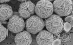
Currently on exhibition as part of the Royal Society’s 350th anniversary celebrations, the Mesolens is a giant microscope that can show large field-of-view images of living specimens in incredible detail – thousands of living cells in focus and in detail at the same time. Until now, scientists have had to rely on low-mag light microscopes to obeserve living specimens, or use sections of dead specimens on an electron microscope in order to get high-mag images.

Mesolens vs Hooke
Robert Hooke first drew a human flea in his 1665 book Micrographia. Along with van Leeuwenhoek, Hooke kick-started microbiology, and so it is a fitting tribute that some 345 years later, LMB give us their flea images. You can pan and zoom across a Mesolens image by clicking here
.
Check out this short article from Wired.com explaining how the Mesolens works, and go to the Laboratory of Molecular Biology’s official site for the Mesolens. Can you distinguish between it and a normal light or electron microscope? What advantages will this give to researchers?
The Guardian has a gallery of images from Mesolens, and there is a short video showing image density from the LMB site, as well as a teachers guide to microscopy.
Cell Theory – Hooke and van Leeuwenhoek
Antonie van Leeuwenhoek invents the microscope:
Rediscovering Robert Hooke (from the Royal Society): Click for video